0
0Chapter 6: Molecular Basis of Inheritance:
Double stranded deoxyribonucleic acid (DNA) acts as genetic material (hereditary molecule) in all cellular organisms (bacteria, protists, fungi, animals and plants). Viruses may have DNA (single stranded DNA- ssDNA or double stranded DNA- dsDNA) or RNA (ssRNA or dsRNA) as their genetic material, but never both. Friedrich Miescher (1844-1895), a Swiss biologist and physician, first isolated DNA from pus of discarded medical bandages in 1869 and called it nuclein because of its presence in the nucleus. Albrecht Kossel (1853-1927), a German biologist and geneticist, isolated five nitrogenous bases (adenine, cytosine, guanine, thymine, and uracil) from nuclein in 1878. Phoebus Levene (1869-1940), an American biochemist, reported DNA and RNA in nuclein. He also found that DNA consists of four nitrogenous bases (adenine, cytosine, guanine, and thymine), a deoxyribose sugar and a phosphate group. Ostwald Avery, Colin Macleod, and Maclyn McCarty found that DNA was the transforming material of Griffith’s transforming experiment in1943. The scientific community did not accept the idea of Avery, MacLeod and McCarty ‘DNA as hereditary molecule’, because some suspected cross-contamination/ impurity of their isolated DNA. Through their experiment on T2 phage in 1952, Alfred Hershey and Martha Chase proved that DNA was the genetic material in the experimental viruses.
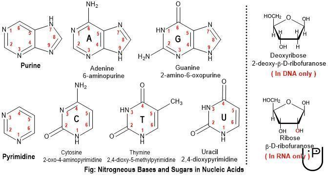
DNA: Deoxyribonucleic Acid: DNA is a heteropolymer of deoxyribonucleotides that consists of a nitrogenous base, a deoxyribose sugar and a phosphate group. Two types of nitrogenous bases are present in DNA. Purine nitrogenous bases are double-ring structure and include adenine (A) and guanine (G). Pyrimidine bases are single-ring structure and include cytosine (C) and thymine (T). The sugar moiety present in DNA is a five-carbon deoxyribose sugar. A nitrogenous base is attached to 1’ carbon with N-glycosidic bond and a phosphate group is esterified to 5’ carbon of the deoxyribose sugar, forming a deoxyribonucleotide (frequently simplified as ‘nucleotide’). A nitrogenous base linked to 1’ carbon of deoxyribose sugar with N-glycosidic bond is called nucleoside (ex- deoxyadenosine, deoxycytosine, deoxyguanosine, deoxythymine). A nucleoside with a phosphate group at 5’ carbon of deoxyribose sugar is called nucleotide (deoxyadenosine monophosphate, deoxycytosine monophosphate, deoxyguanosine monophosphate, deoxythymine monophosphate).
The –OH group at 5’ carbon of the sugar of a nucleotide forms a covalent bond with an oxygen atom in the phosphate group of adjacent nucleotide following the removal of a water molecule (condensation). A nucleotide (say, first nucleotide) binds to adjacent nucleotide (say, second nucleotide) with 3’-5’- phosphodiester bond.
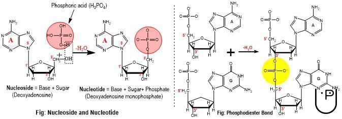
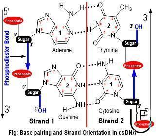
A phosphate group (ionized phosphoric acid, H3PO4) links 3’-OH of first nucleotide to 5’-OH of next nucleotide though ester bond with each –OH group, thus called 3’-5’ phosphodiester bond. The linear polymer of nucleotides linked by adjacent phosphodiester bonds is called a strand. Thus, phosphodiester bonds also act as the backbone of DNA strand. Each strand has a terminal (at one end, not free to make any bond) phosphate group linked to 5’C of deoxyribose sugar of first nucleotide, termed as 5’ end of DNA stand. The last nucleotide of the strand has a free 3’- OH in the deoxyribose sugar to which new nucleotides can be added. This end with free 3’-OH group is called 3’ end of DNA strand. Thus, each DNA strand has a 5’-3’ polarity i.e. it extends from 5’ end to 3’ end. In a double stranded DNA (DNA with two strands of DNA), the two strands run antiparallel to each other. At either end of the dsDNA, one strand has a terminal 5’-phosphate and the other strand has a free 3’-OH group, or vice-versa. Both the strands are complementary to each other. Adenine on one strand forms two hydrogen bonds with thymine located oppositely on the other strand. Likewise, guanine on one strand forms three hydrogen bonds with cytosine located oppositely on the other strand. Two nucleotides joined together by hydrogen bonds form a base pair (ex- A-T base pair and G-C base pair). Hydrogen bonds formed among oppositely placed nucleotides on the two strands stabilizes the double stranded structure of DNA.
Chargaff’s Rules: Erwin Chargaff (1905-2002), an Austrian biochemist, proposed two rules while working on the base composition of DNA. The first rule states that in a double stranded DNA, the number purine bases is equal to pyrimidine bases. Alternatively, adenine residues is equal to that of thymine and number of guanine residues is equal to that of cytosine. The second rule states that the relative amount of A, C, T and G residues varies from species to species. Chargaff also discussed his ideas to Watson and Crick in 1952 at Cambridge.
Watson & Crick Model of DNA: Rosalind E Franklin (1920-1958) first presented the X-ray diffraction pattern of A and B DNA in 1951- Watson was one of the attendees of the seminar. She and Raymond Gosling (then, Franklin’s Ph.D student) also obtained another X-ray diffraction of B-DNA with better resolution in 1952. This photograph taken by Gosling is called ‘photograph 51’. The photograph on X-ray diffraction pattern of DNA depicts that the phosphate atoms are located at the periphery and DNA has water molecules associated with it. The regular ‘X’ pattern of diffraction in her photograph also suggests that B-DNA takes a regular right-handed helix configuration. When asked by Franklin, Gosling gave the photograph to Maurice Wilkins in 1952. Without taking any consent from Franklin, Wilkins shared the date with Watson and Crick.
Considering the data from X-ray diffraction patterns and base pairing behavior of DNA, James D Watson and Francis Crick proposed double helix structure of DNA in 1953. Watson, Crick and Maurice HF Wilkins got the Nobel Prize for the year 1962 in medicine or physiology.
The important points of Watson and Crick model of DNA are as follow:
- DNA is composed of two antiparallel strands held together by hydrogen bonds between complementary nitrogenous bases of the nucleotides placed oppositely on the two strands. Because adenine (A) always pairs with thymine (T) and guanine (G) always pairs with cytosine (C), the base pairing is complementary. The complementary base pair also results the two strands to be complementary of each other. If the base sequence of one strand is known, the base sequence of the complementary strand can easily be depicted. The complementarity of strands ensures DNA functioning well as the genetic material as well as also aid in DNA repair or other events.
- 5’-3’ phosphodiester bond acts as the backbone of each DNA strand. The phosphodiester backbone lies at the periphery of the helix and the nucleotides are projected towards the central axis of the helix.
- The phosphate groups provide an overall negative charge to a DNA molecule.
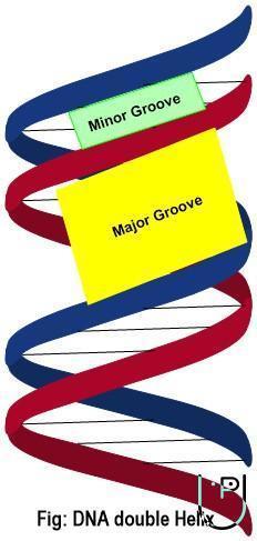
- The two strands of DNA wind around each other forming a right handed helix. The right helix sense means that when the helix is look from the top, the helix follows a clockwise path and moves away from the observer. One complete turn of the helix (3600) of DNA has approximately 10 base pairs.
- The diameter of the helix i.e. the distance between phosphate atom at the backbone and the central axis, is 2 nanometers (20 angstroms, Å). The constancy of 2 nm helix diameter along the helix length lies in the fact that a pyrimidine (single ring) on one strand always pairs with a purine (double ring) on the opposite strand. The adjacent base pairs on the dsDNA helix are 0.34 nm apart along the longitudinal axis while taking the helical path. This distance is called rise per base pair (0.34 nm= 3.4 Å). The distance along the central longitudinal axis for a complete helix turn, called the pitch of the helix, is 3.4 nm (= 34 Å).
- The bases are placed approximately perpendicular to the central axis. The base staking is stabilizing by hydrophobic interactions and van der Waal forces between the adjacent bases.
- The nitrogen atom linked to carbon 6 of adenine and carbon 4 of cytosine predominantly take amino (-NH2) configuration rather than the imino (=NH) form. The oxygen atom linked to carbon 6 of guanine and carbon 4 of thymine predominantly take keto (-C=O) configuration rather than the enol (-C-OH) form. These configurations ensure that adenine exclusively pairs with thymine and guanine exclusively pairs with cytosine.
- The spaces between adjacent turns of the helix form two types of grooves with different width. The groove with relatively greater width is called major groove. The groove with relatively narrower width is called minor groove. Many DNA binding proteins bind to better-exposed specific base sequences of the major groove and carry out the respective catalysis. Polymerases (DNA/ RNA polymerases), nucleases, transcription factors, histones, etc. interact with DNA at its major groove. The DNA binding proteins may possess zinc finger, leucine zipper, helix-turn-helix and/or many other domains that interacts to specific base sequence at the major groove. Few DNA binding proteins like netrospin, distamycin, etc. interact with the specific base sequences of the minor groove.
Deviations from Watson - Crick Model:
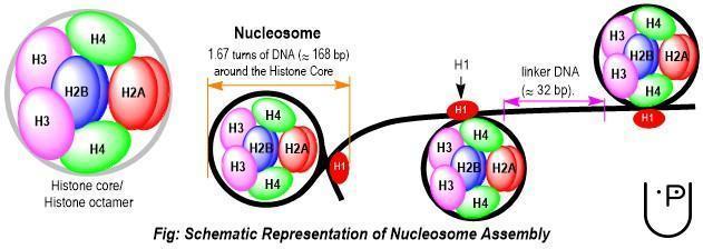
DNA Supercoiling: Each eukaryotic chromosome has a single dsDNA associated with several proteins, which are crucial for its stability, replication, transcription and other functions. A DNA molecule with its associated histone proteins and RNA is called chromatin. Histones are a family of small, positively charged (because of being rich in lysine and arginine amino acids) proteins. The positive charge on histones provides thermodynamically stable interactions with negatively charged DNA. The interaction between histones and DNA is purely non-covalent in nature that ensures their assembly and disassembly during cellular processes like chromosome condensation/ decondensation, DNA replication, transcription or in response to other cell signaling events. Nucleosome is the lowest level of organization of DNA into chromosome. These are also the repeating, structural and functional units of chromosome. A nucleosome consists of around 146 base pairs of DNA taking about 1.67 turns around the central histone octamer core. The histone octamer core consists of four heterodimers- two (H2A-H2B) and two (H3-H4) dimers at its center. A molecule of H1 seals the association of 1.67 turns of DNA around the histone core. The total length of DNA interacting with a nucleosome and a H1 associated to it is 168 base pairs. H1 histone also interacts with other histones during condensation of DNA into chromosome. When chromatin fiber is treated with a low ionic strength solution, H1 histones dissociate from it. The resultant structure consists of the nucleosomes adjacently linked together by linker DNA in between them, and appears like ‘beads on strings’ under electron microscope. The length of DNA joining two adjacent nucleosomes is called linker DNA (≈ 22 bp).
Nucleosome has a diameter of ≈10 nm, thus this arrangement of DNA into nucleosome is also called 10 nm fiber. Nucleosome wraps DNA around it with a packing ratio of approximately 7:1 meaning that nucleosome (dia= 10 nm) wraps about 7 times longer DNA strand (≈ 70 nm) around it; or alternatively nucleosome packing reduces the overall length of DNA strand seven folds.
In next higher level of organization, histones of one nucleosome interact with adjacent ones to form 30 nm fiber with a packing ration of ≈ 40:1. In 30 nm fiber, N-terminal tail of H4 histones of one nucleosome interacts with H1 and H2A/H2B histones of adjacent nucleosomes and causes packing of nucleosomes in 30 nm fiber. 30 nm fiber formation occurs through debated zigzag or solenoid model of nucleosome arrangement. The metaphase chromosome has a packing ration of 10,000:1 in which 30 nm fibers form supercoiled loops (stabilized by cohesin ring) and bound to a structural framework/ scaffold consisting of condensin multiprotein complexes.
Heterochromatin & Euchromatin: The relatively compact and transcriptionally inactive parts of an interphase chromosome are called heterochromatin. Constitutive heterochromatins remain transcriptionally inactive and highly condensed in all types of cells all the time. Such permanently inactive regions constitute approximately 10% of a eukaryotic chromosome and are generally present in telomeres and centromeres of the chromosome. Facultative heterochromatins are type of heterochromatin that remain inactive at some stage and become active at another stage of life cycle. For example, the genes expressed at and after puberty constitute facultative heterochromatin.
Griffith’s Experiment: Fredrick Griffith, an English physician, first reported transformation of one strain of Streptococcus pneumoniae into other strain in 1928. The bacteria cause pneumonia. A strain of this bacterium produces capsule and gives a smooth, shiny colony on culture media, thus called S-strain (smooth strain). The S-strain exhibits pathogenicity in laboratory mice. Another strain without capsule produces rough and coarse colony on culture medium, thus called R-strain (rough strain). The R-strain is avirulent (non-pathogenic).
| Mice injected with | Outcome | |
| 1 | Live S-strain (virulent) | Pneumonia → mice dead |
| 2 | Live R-strain (avirulent) | No pneumonia → mice alive |
| 3 | Heat killed S-strain + live R-strain | Pneumonia → mice dead |
When injected with live S-strain bacteria, mice developed pneumonia and died. When injected with live R-strain bacteria, mice did not develop pneumonia and survived. When heat killed S-strain injected to mice, they remain unaffected. However, when heat killed S-strain bacteria mixed with live R-strain injected to mice, they developed pneumonia and died. He was also able to isolate and culture S-strain bacteria from affected mice. Since heat killed bacteria could have no way been revived, he concluded that some molecules from heat killed S-strain had transformed the R-strain into S-train.
Avery - McLeod - McCarty Experiment: In 1944, Oswald Avery, Colin McLeod and Maclyn McCarty carried out Griffith’s experiment at molecular level in search of the S-strain’s biomolecule that transforms R-strain. They heat killed S-strain bacteria and prepared its homogenate in saline. The homogenate is treated with physical or chemical agents and hydrolyti