


Your Worksheet is Ready
CBSE - Class 11 Biology Photosynthesis Worksheet
EXERCISES
Figure 11.10 shows the effect of light on the rate of photosynthesis. Based on the graph, answer the following questions: (a) At which point/s (A, B or C) in the curve light is a limiting factor?

Figure 11.10 shows the effect of light on the rate of photosynthesis. Based on the graph, answer the following questions: (b) What could be the limiting factor/s in region A?
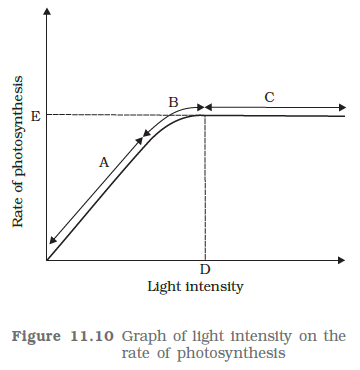
Figure 11.10 shows the effect of light on the rate of photosynthesis. Based on the graph, answer the following questions: (c) What do C and D represent on the curve?
Worksheet Answers
Solution:
The green color of a leaf is primarily attributed to the presence of Chlorophyll ($a$ and $b$), which are localized within the thylakoid membranes of chloroplasts. These pigments absorb light energy for photosynthesis [Per the Light-Dependent Reactions of Photosynthesis]. When a leaf is kept in the dark, the following physiological shifts occur:
To determine the relative stability, consider the following comparative data:
| Pigment | Chemical Nature | Relative Stability |
|---|---|---|
| Chlorophyll $a/b$ | Porphyrin ring with Magnesium center | Low (Light-dependent degradation) |
| Carotenoids | Tetraterpenoid hydrocarbons | High (Light-independent stability) |
Conclusion on Stability: Carotenoids are significantly more stable than chlorophyll. While chlorophyll is highly reactive and susceptible to photo-oxidation and enzymatic degradation, carotenoids function as photoprotective agents that dissipate excess energy and maintain structural integrity even under prolonged dark conditions [Per the Accessory Pigment Photoprotection Hypothesis].
The yellowing (chlorosis) occurs because chlorophyll is metabolically expensive and unstable in the dark, leading to its rapid catabolism. Carotenoids are inherently more stable, acting as a secondary pigment profile that persists after the primary photosynthetic pigments have decomposed. This shift is a survival mechanism to minimize metabolic demand when energy gain from light is unavailable.
Final Solution: The leaf turns yellow because chlorophyll degrades in the dark due to its instability and the cessation of light-dependent synthesis. Carotenoids are significantly more stable than chlorophyll, as they do not require light for structural maintenance and serve as a persistent pigment pool in the absence of chlorophyll.
Solution:
Photosynthesis is dependent on the existence of a Reaction Center within the Photosystems (PS I and PS II). The core of this reaction center consists of specialized molecules of Chlorophyll $a$ ($P_{700}$ in PS I and $P_{680}$ in PS II).
Plants utilize accessory pigments, including Chlorophyll $b$, carotenoids, and xanthophylls, for the following biological imperatives:
| Function | Biological Justification |
|---|---|
| Broadening Absorption Spectrum | Chlorophyll $a$ absorbs primarily in the blue-violet and red regions. Accessory pigments fill the "green gap," capturing light energy in wavelengths (450–550 nm) that Chlorophyll $a$ cannot effectively utilize. [Per the Action Spectrum of Photosynthesis]. |
| Photoprotection | High-intensity light can cause the formation of reactive oxygen species (ROS) or triplet-state chlorophyll, which are damaging to the thylakoid membranes. Carotenoids absorb excess energy and dissipate it as heat, preventing photo-oxidative damage. |
| Energy Transfer Efficiency | Accessory pigments act as a "funnel" (Antenna Complex), channeling harvested photons toward the Reaction Center ($P_{680}/P_{700}$) to ensure maximum rate of reaction even in low-light conditions. |
The system is hierarchical. The accessory pigments ($Chl\ b$, Carotenoids) are evolutionarily conserved because they act as light-harvesting antennas that increase the cross-sectional area for photon capture. Without the core $Chl\ a$ molecule, the conversion of light energy to chemical potential energy ($ATP$ and $NADPH$) cannot initiate, regardless of the quantity of accessory pigments present. Thus, $Chl\ a$ is the reaction center pigment, while $Chl\ b$ and others are antenna pigments that broaden the utilization of the solar spectrum and protect the photosystem from excitation damage.
Final Solution: No, the plant would not carry out photosynthesis because Chlorophyll $a$ is the essential pigment required to initiate the photochemical reaction (charge separation). Accessory pigments, including Chlorophyll $b$, are present to broaden the absorption spectrum and provide photoprotection, enhancing the overall efficiency and survival of the photosynthetic apparatus.
Solution:
When comparing leaves from a sunny environment versus a shady environment, empirical observation reveals that leaves grown in the shade exhibit a darker green coloration compared to those exposed to direct, high-intensity sunlight. This phenomenon is a physiological adaptation driven by the necessity to maximize photosynthetic efficiency under light-limiting conditions.
The intensity of green coloration in a leaf is directly proportional to the density of chlorophyll pigments within the mesophyll cells [Per the Beer-Lambert Law application in biological systems].
| Characteristic | Sun-Grown Leaf | Shade-Grown Leaf |
|---|---|---|
| Chlorophyll Content | Lower | Higher |
| Leaf Color | Light Green | Dark Green |
| Photosynthetic Adaptation | High light saturation capacity | High light-harvesting efficiency |
The darker green color in shade leaves is a morphological and biochemical response known as shade-induced greening. By increasing the chlorophyll content, the plant optimizes its ability to harvest the limited quanta of light available in shaded environments, ensuring that the light-dependent reactions of photosynthesis can proceed at a rate sufficient to maintain metabolic homeostasis [Justified by the Law of Limiting Factors, Blackman, 1905].
Final Solution: Leaves grown in the shade are darker green because they possess a higher density of chlorophyll pigments, which allows the plant to maximize photon capture in light-deficient environments.
Solution:
Determining the photosynthetic pathway ($C_3$ or $C_4$) of a plant solely by its external morphology is generally not possible with absolute certainty. While certain physiological traits often correlate with $C_4$ photosynthesis, external appearance is not a diagnostic characteristic. The distinction is fundamentally anatomical and biochemical, occurring at the cellular and molecular levels.
External morphology (leaf shape, size, color, or phyllotaxy) is a result of evolutionary adaptation to environmental pressures such as water availability, light intensity, and herbivory, rather than a direct indicator of the carbon fixation mechanism. Both $C_3$ and $C_4$ plants can exhibit identical growth forms (e.g., both groups contain various species of grasses and herbs).
The primary diagnostic feature is Kranz Anatomy, which is characteristic of $C_4$ plants but absent in $C_3$ plants. This requires internal examination of a leaf cross-section via microscopy.
| Feature | $C_3$ Plant | $C_4$ Plant |
|---|---|---|
| Bundle Sheath Cells | Small, thin-walled, non-photosynthetic | Large, thick-walled, rich in chloroplasts |
| Leaf Anatomy | Typical mesophyll organization | Kranz anatomy (Wreath-like arrangement) |
| Chloroplasts | Uniform in mesophyll cells | Dimorphic (Mesophyll: Granal; Bundle Sheath: Agranal) |
To definitively identify the pathway, one must perform internal analysis:
Final Solution: It is impossible to identify a plant as $C_3$ or $C_4$ solely by external inspection. This distinction is based on internal leaf anatomy (Kranz anatomy) and biochemical differences (primary carboxylating enzymes), which are only visible via microscopic examination and biochemical assays.
Solution:
| Feature | Cyclic Photophosphorylation | Non-Cyclic Photophosphorylation |
|---|---|---|
| Photosystems Involved | Only Photosystem I (PSI / $P_{700}$) | Both Photosystem II ($P_{680}$) and Photosystem I ($P_{700}$) |
| Products Formed | Only $ATP$ | $ATP$, $NADPH + H^+$, and $O_2$ (byproduct) |
| Electron Source | The excited reaction center ($P_{700}$) itself | Photolysis of Water ($H_2O$) |
| Electron Pathway | Cyclic (returns to $P_{700}$) | Z-scheme (non-cyclic) |
| Location | Stroma lamellae | Thylakoid membranes (Grana) |
In cyclic photophosphorylation, the electron emitted by $P_{700}$ is captured by the primary electron acceptor and passed through a series of carriers: Ferredoxin ($Fd$), Cytochrome $b_6f$ complex, and Plastocyanin ($Pc$). [Per the Chemiosmotic Hypothesis]. As electrons move through the Cytochrome complex, protons ($H^+$) are pumped into the lumen, generating a proton gradient that drives $ATP$ synthase. The electron eventually returns to the reaction center ($P_{700}$), hence completing a cycle. No $NADPH$ is produced.
In non-cyclic photophosphorylation, the Z-scheme occurs. $PSII$ ($P_{680}$) absorbs light, loses an electron to the primary acceptor, and replaces it via the photolysis of water: $2H_2O \rightarrow 4H^+ + O_2 + 4e^-$. [By the Photolysis Law]. The electrons pass from $PSII$ through Plastoquinone ($PQ$), Cytochrome $b_6f$, and Plastocyanin to reach $PSI$. Subsequently, $PSI$ is energized and transfers electrons to $NADP^+$ via Ferredoxin to produce $NADPH$.
Final Solution: Cyclic photophosphorylation is restricted to PSI and results solely in ATP synthesis to meet cellular energy demands, while non-cyclic photophosphorylation utilizes both PSI and PSII to produce both ATP and NADPH while facilitating the photolysis of water.
Solution:
The fundamental distinction in leaf anatomy between $C_3$ and $C_4$ plants lies in the spatial organization of photosynthetic tissues. While $C_3$ plants utilize a standard mesophyll arrangement, $C_4$ plants have evolved a specialized morphology known as Kranz Anatomy [Per the Evolutionary Adaptations for Photorespiration Suppression].
| Characteristic | $C_3$ Leaf Anatomy | $C_4$ Leaf Anatomy |
|---|---|---|
| Mesophyll Differentiation | Differentiated into palisade and spongy parenchyma. | Undifferentiated; cells are radially arranged around the bundle sheath. |
| Bundle Sheath Cells | Small, thin-walled, and lack chloroplasts or contain few. | Large, thick-walled, and contain numerous prominent chloroplasts. |
| Kranz Anatomy | Absent. | Present (Wreath-like arrangement of mesophyll). |
| Chloroplast Dimorphism | Absent (All chloroplasts are structurally similar). | Present (Mesophyll chloroplasts are granal; Bundle sheath chloroplasts are agranal). |
In $C_4$ anatomy, the Kranz anatomy serves to spatially separate the initial $CO_2$ fixation and the Calvin cycle. The radial arrangement of mesophyll cells allows for high-efficiency capture of $CO_2$ by Phosphoenolpyruvate (PEP) carboxylase [Per the C4 Cycle mechanism]. The large, thick-walled bundle sheath cells, containing agranal chloroplasts, maintain high internal $CO_2$ concentrations, which effectively inhibits the oxygenase activity of RuBisCO, thereby suppressing photorespiration [Justified by the lack of direct $O_2$ access in the sheath]. Conversely, in $C_3$ plants, mesophyll cells perform all light-dependent and dark reactions in a single cell type, rendering them susceptible to photorespiratory losses when temperatures rise.
Final Solution: The fundamental anatomical difference is the presence of Kranz anatomy in $C_4$ plants (radially arranged mesophyll around large, chloroplast-rich bundle sheath cells), which is absent in $C_3$ plants, facilitating specialized spatial separation of the photosynthetic pathway.
Solution:
The high productivity of $C_4$ plants (e.g., maize, sugarcane) compared to $C_3$ plants, despite the localization of the Calvin cycle solely to bundle sheath cells, is attributed to the spatial separation of carbon fixation and the elimination of photorespiration. This is facilitated by the Kranz anatomy.
In $C_4$ plants, $CO_2$ fixation occurs in two distinct cell types. Atmospheric $CO_2$ is first fixed in the mesophyll cells by Phosphoenolpyruvate (PEP) carboxylase. [Per the Hatch-Slack pathway]. Unlike RuBisCO, PEP carboxylase has a very high affinity for $HCO_3^-$ and lacks oxygenase activity, ensuring rapid carbon capture even at low $CO_2$ concentrations.
The four-carbon acid (malate/aspartate) is transported to the bundle sheath cells, where it undergoes decarboxylation. This release of $CO_2$ creates a high-concentration environment around the RuBisCO enzyme. [By the Principle of Competitive Inhibition], high levels of $CO_2$ outcompete $O_2$ for the active site of RuBisCO, effectively suppressing photorespiration—a wasteful process in $C_3$ plants that consumes ATP and releases previously fixed carbon.
| Feature | $C_3$ Plants | $C_4$ Plants |
|---|---|---|
| Primary CO2 Acceptor | RuBP | PEP |
| Photorespiration | High | Negligible |
| Productivity | Moderate | High |
Final Solution: $C_4$ plants are highly productive because they utilize the Kranz anatomy to concentrate $CO_2$ within bundle sheath cells, effectively neutralizing the oxygenase activity of RuBisCO and eliminating the loss of fixed carbon through photorespiration.
Solution:
The distinction between $C_3$ and $C_4$ plants is fundamentally determined by the leaf's internal anatomical organization. Specifically, the presence of Kranz Anatomy is the diagnostic structural feature used to classify a plant as a $C_4$ plant.
In $C_4$ plants (e.g., maize, sugarcane), the leaves exhibit a specialized arrangement of cells known as "Kranz" anatomy (German for "wreath"). This structure serves to spatially separate the light-dependent reactions and the Calvin cycle to minimize photorespiration.
| Feature | $C_3$ Plant Anatomy | $C_4$ Plant Anatomy |
|---|---|---|
| Bundle Sheath | Small, lack chloroplasts or have reduced ones. | Large, contain numerous, starch-rich chloroplasts. |
| Mesophyll Arrangement | Differentiated into palisade and spongy parenchyma. | Undifferentiated, arranged radially around bundles. |
| Photorespiration | Significant (due to RuBisCO oxygenation). | Negligible (due to high $CO_2$ concentration). |
The structural differentiation described above facilitates the C4 cycle [Per the Hatch-Slack Pathway]:
If a cross-section of the leaf shows distinct, large-celled bundle sheaths packed with chloroplasts and a radial arrangement of mesophyll, it is a $C_4$ plant. The absence of such organization, where mesophyll is loosely arranged and bundle sheath cells are minimal/chloroplast-deficient, characterizes a $C_3$ plant.
Final Solution: The presence or absence of "Kranz Anatomy" (specifically the presence of large, chloroplast-rich bundle sheath cells arranged in a wreath-like layer around vascular bundles) allows for the definitive identification of a $C_4$ plant versus a $C_3$ plant.
Solution:
The rate of photosynthesis is governed by the Law of Limiting Factors, which states that if a chemical process is affected by more than one factor, its rate is determined by the factor that is nearest to its minimal value [Per Blackman's Principle, 1905]. In the context of light intensity and photosynthesis, light acts as a limiting factor when its scarcity restricts the conversion of light energy into chemical energy, thereby capping the production of NADPH and ATP required for the Calvin Cycle.
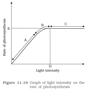
The graph represents the Rate of Photosynthesis ($y$-axis) against Light Intensity ($x$-axis).
To determine where light is the limiting factor, we apply the mathematical derivative of the rate ($R$) with respect to light intensity ($I$):
$\frac{dR}{dI} > 0$ (Rate is limited by $I$)
$\frac{dR}{dI} \approx 0$ (Rate is limited by other factors, $I$ is in excess)
Based on the gradient analysis:
| Point | Status | Reasoning |
|---|---|---|
| A | Limiting | The rate is in the linear phase; increasing light intensity directly increases the rate. |
| B | Transition | The rate is approaching saturation; light influence is diminishing as internal metabolic limits emerge. |
| C | Non-limiting | Saturation point; photosynthesis is limited by other factors (e.g., substrate concentration, enzyme kinetics). |
Final Solution: Light is a limiting factor at points A and B, as these segments demonstrate a direct dependence of the photosynthetic rate on light intensity. Point C represents the light saturation point where light is no longer the limiting factor.
Solution:
In the standard representation of the light-response curve of photosynthesis, the graph plots the Rate of Photosynthesis (y-axis) against Light Intensity (x-axis). Region A represents the initial, linear portion of the curve where the photosynthetic rate increases proportionally with light intensity.
Per Blackman’s Law of Limiting Factors (1905), if a chemical process is affected by more than one factor, then its rate will be determined by the factor which is nearest to its minimal value; it is the factor which directly affects the process if its quantity is changed.
In Region A, the rate of photosynthesis is strictly dependent on the light intensity. As light intensity increases, the rate of production of $ATP$ and $NADPH$ (the products of the light-dependent reactions) increases proportionally. Consequently, the rate of $CO_2$ fixation is directly limited by the availability of these light-generated products [Per the Light-Dependent Reaction stoichiometry].
The linearity of the graph in Region A indicates that the system has not yet reached light saturation. Therefore, any increment in light intensity results in a proportional increment in the photosynthetic rate. If the rate were limited by other factors (such as $CO_2$ or enzymes), the curve would deviate from this linear trajectory before the light saturation point is reached.
Final Solution: The limiting factor in region A is Light Intensity. As the light intensity increases, the rate of photosynthesis increases linearly, confirming that light is the factor nearest to its minimum value per Blackman's Law of Limiting Factors.
Solution:
The relationship between light intensity and the rate of photosynthesis is characterized by a non-linear trajectory. Initially, as light intensity increases, the rate of photosynthesis increases linearly because light energy is the limiting factor [Per the Law of Limiting Factors by Blackman, 1905]. As intensity continues to rise, the rate becomes limited by other environmental factors (e.g., $CO_2$ concentration or temperature), resulting in a plateau.
Point C represents the stage where the rate of photosynthesis begins to deviate from a linear progression. At this intensity, light is no longer the sole limiting factor. Internal cellular processes or the concentration of $CO_2$ start to influence the rate of carbon fixation [Justification: Principles of Physiological Ecology]. The curve at Point C is in a state of diminishing returns.
Point D corresponds to the Light Saturation Point. At this specific intensity, the rate of photosynthesis reaches a maximum plateau ($P_{max}$). Beyond this value, increasing the light intensity further yields no significant increase in the rate of photosynthesis because the light-independent reactions (Calvin Cycle) or the availability of enzymes (e.g., RuBisCO) and substrates become the absolute limiting factors [Per the Law of Limiting Factors].
| Feature | Point C | Point D |
|---|---|---|
| Definition | Transition phase | Saturation point |
| Limiting Factor | Light is co-limiting | Other factors (CO2/Temperature) |
| Rate Status | Increasing at a decreasing rate | Maximum constant rate |
Final Solution: Point C represents the stage of transition where light intensity starts to become saturated and other factors begin to limit the rate; Point D represents the light saturation point where the rate of photosynthesis reaches its maximum and remains constant despite further increases in light intensity.
Solution:
The photosynthetic process is bifurcated based on the initial carbon fixation product and the physiological adaptation of the plant species to varying environmental conditions.
| Feature | $C_3$ Pathway (Calvin Cycle) | $C_4$ Pathway (Hatch-Slack Pathway) |
|---|---|---|
| Primary CO$_2$ Acceptor | Ribulose-1,5-bisphosphate (RuBP) | Phosphoenolpyruvate (PEP) |
| First Stable Product | 3-phosphoglycerate (3-PGA) | Oxaloacetate (OAA) |
| Carbon Fixation Enzyme | RuBisCO only | PEP Carboxylase (primary), RuBisCO (secondary) |
| Anatomy | Standard mesophyll cell structure | Kranz anatomy (Mesophyll + Bundle Sheath cells) |
| Photorespiration | High | Negligible |
| Optimum Temperature | $20^\circ\text{C} - 25^\circ\text{C}$ | $30^\circ\text{C} - 45^\circ\text{C}$ |
The $C_4$ pathway is distinguished by its spatial separation of fixation and reduction, which facilitates the concentration of CO$_2$ around RuBisCO, thereby minimizing the oxygenase activity of the enzyme [Per the Law of Competitive Inhibition].
While $C_3$ plants are efficient in temperate environments, $C_4$ plants require extra ATP to regenerate PEP. However, because $C_4$ plants effectively pump CO$_2$ into the bundle sheath cells, they suppress photorespiration—a wasteful process where RuBisCO acts as an oxygenase [Per the RuBisCO Oxygenase vs. Carboxylase kinetics].
Final Solution: The primary difference lies in the anatomical segregation of reactions in $C_4$ plants (Kranz anatomy) compared to the single-cell fixation in $C_3$ plants, allowing $C_4$ species to significantly minimize photorespiratory losses.
Solution:
Ribulose-1,5-bisphosphate carboxylase-oxygenase (RuBisCO) is the most abundant enzyme on Earth. Its catalytic activity is governed by the relative concentrations of $CO_2$ and $O_2$ at the active site, as well as the temperature. The dual nature is defined by the following reactions:
The oxygenation process is metabolically wasteful, as it releases fixed carbon and consumes ATP. $C_4$ plants have evolved a specialized mechanism to bypass this inefficiency.
Unlike $C_3$ plants, $C_4$ plants possess Kranz Anatomy, which spatially separates the primary $CO_2$ fixation from the Calvin Cycle. This separation is the prerequisite for increasing the $CO_2:O_2$ ratio at the site of RuBisCO.
In $C_4$ plants, the initial fixation of $CO_2$ occurs in the mesophyll cells, while the Calvin Cycle (involving RuBisCO) is restricted to the bundle sheath cells.
The bundle sheath cells in $C_4$ plants are characterized by:
By concentrating $CO_2$ in the bundle sheath and simultaneously minimizing the presence of $O_2$, the $C_4$ cycle ensures that the probability of RuBisCO binding $O_2$ is virtually zero. Consequently, the enzyme is "forced" to function as a carboxylase. This adaptation prevents the energy-intensive process of photorespiration, thereby significantly increasing the photosynthetic efficiency of the plant under high-light and high-temperature conditions.
Final Solution: RuBisCO carries out more carboxylation in $C_4$ plants because they utilize a $CO_2$ concentrating mechanism that elevates the local concentration of $CO_2$ around the enzyme while excluding $O_2$ within the bundle sheath cells, thereby suppressing the oxygenase activity of RuBisCO.