


Your Worksheet is Ready
CBSE - Class 9 Science Tissues Worksheet
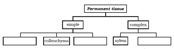
Complete the following chart:
Worksheet Answers
Solution:
The muscular tissue in vertebrates is categorized into three distinct types based on their histological characteristics and physiological roles. The following table provides the precise differentiation required for academic mastery:
| Feature | Striated (Skeletal) Muscle | Unstriated (Smooth) Muscle | Cardiac Muscle |
|---|---|---|---|
| Cell Shape | Cylindrical, unbranched. | Spindle-shaped (fusiform). | Cylindrical, branched. |
| Nuclei | Multinucleated (Peripheral). | Uninucleated (Central). | Uninucleated (Central). |
| Striations | Present (Light/Dark bands). | Absent (Smooth appearance). | Present (Faint striations). |
| Control | Voluntary (Somatic nervous system). | Involuntary (Autonomic nervous system). | Involuntary (Autonomic nervous system). |
| Location | Attached to bones (Skeletal system). | Walls of hollow organs (e.g., gut, blood vessels). | Walls of the heart (Myocardium). |
A. Striated Muscles [Per Physiological Function]: These cells possess alternating light and dark bands (actin and myosin filaments), creating the "striated" effect. Their attachment to the skeleton via tendons allows for rapid, voluntary movement of limbs [Ref: Sarcomere Contraction Theory].
B. Unstriated (Smooth) Muscles [Per Homeostatic Regulation]: These lack the protein arrangement that produces striations. They exhibit slow, rhythmic contractions known as peristalsis, essential for involuntary internal processes like digestion and blood pressure regulation [Ref: Autonomic Nervous Control].
C. Cardiac Muscles [Per Specialized Endurance]: These are unique because they are involuntary like smooth muscles but possess striations like skeletal muscles. The presence of intercalated discs between cells allows for the transmission of electrical impulses as a single unit (syncytium), ensuring the heart pumps rhythmically throughout an individual's lifespan without fatigue [Ref: Electrophysiological Coupling].
Final Solution: The three muscle types differ fundamentally in cellular morphology (cylindrical vs. spindle-shaped), the presence or absence of striations (actin-myosin overlap patterns), and their physiological sites (skeletal attachments vs. visceral organs vs. myocardium).
Solution:
The neuron (nerve cell) is the structural and functional unit of the nervous system. It is specialized to receive, process, and transmit information via electrical and chemical signals [Per the Electrophysiological Principle of Cellular Communication].
| Structure | Primary Function |
|---|---|
| Dendrite | Signal Reception (Afferent) |
| Cyton | Integration & Protein Synthesis |
| Axon | Signal Conduction (Efferent) |
Final Solution: The neuron consists of the cyton, dendrites, and a myelinated axon, functioning collectively as a biological relay system to transmit electrochemical impulses across the nervous system.
Solution:
The tissue responsible for the storage of fat in the human body is identified as Adipose tissue. [Per histological classification of connective tissues].
Adipose tissue is a specialized form of loose connective tissue. Its primary biological function is the storage of lipids (triglycerides) within specialized cells known as adipocytes.
| Feature | Description |
|---|---|
| Tissue Type | Loose Connective Tissue |
| Primary Cell Type | Adipocytes |
| Stored Substance | $C_{57}H_{110}O_6$ (Typical Triglyceride) |
Final Solution: The tissue that stores fat in the body is Adipose Tissue.
Solution:
Xylem is a complex permanent tissue responsible for the upward conduction of water and dissolved minerals from the roots to the other parts of the plant. Being a "complex" tissue, it is composed of more than one type of cell working together as a single unit to perform a common function [Per the biological definition of complex tissues].
The xylem tissue is composed of four distinct types of elements, known as xylem elements. These components vary in their structure, function, and presence of protoplasm.
| Element Name | Functional Characteristic |
|---|---|
| Xylem Tracheids | Elongated, tube-like cells with thick, lignified walls; dead at maturity. |
| Xylem Vessels | Long, cylindrical, pipe-like structures; dead at maturity. |
| Xylem Parenchyma | Living cells that store food and assist in the lateral conduction of water. |
| Xylem Fibers (Sclerenchyma) | Dead, sclerified cells providing mechanical support to the plant. |
Final Solution: Xylem tissue is composed of 4 types of elements: Xylem Tracheids, Xylem Vessels, Xylem Parenchyma, and Xylem Fibers.
Solution:
In plant biology, tissues are categorized based on the uniformity and functional specialization of the cells comprising them. The fundamental distinction lies in cell composition and organizational complexity.
| Feature | Simple Permanent Tissue | Complex Permanent Tissue |
|---|---|---|
| Composition | Composed of only one type of cell [Uniform structure]. | Composed of more than one type of cell [Heterogeneous structure]. |
| Primary Function | Storage, photosynthesis, and structural support. | Conduction of water/nutrients and structural reinforcement. |
| Cell Types | Parenchyma, Collenchyma, Sclerenchyma. | Xylem (Tracheids, Vessels, Xylem Fibres, Xylem Parenchyma) and Phloem (Sieve Tubes, Companion Cells, Phloem Fibres, Phloem Parenchyma). |
| Evolutionary Role | Basic structural framework of the plant body. | Advanced vascular system for long-distance transport. |
Final Solution: Simple tissues are groups of structurally and functionally identical cells that perform singular tasks, whereas complex tissues consist of multiple distinct cell types that function as a coordinated unit to facilitate high-order physiological processes such as vascular transport.
Solution:
The fundamental structural differences between parenchyma, collenchyma, and sclerenchyma tissues are primarily dictated by the chemical composition, deposition patterns, and thickness of their respective cell walls.
| Tissue Type | Cell Wall Characteristics | Chemical Composition |
|---|---|---|
| Parenchyma | Thin, uniform, and flexible cell walls. | Primary wall composed mainly of cellulose and pectin. |
| Collenchyma | Irregularly thickened at the corners (angles). | Cellulose, hemicellulose, and high concentrations of pectin. |
| Sclerenchyma | Extremely thick, rigid, and lignified walls. | Secondary wall heavily impregnated with lignin [Per the principle of secondary wall thickening]. |
Final Solution: Parenchyma possesses thin primary walls of cellulose; collenchyma exhibits pectin-rich irregular thickening at corners for flexibility; sclerenchyma features heavily lignified, rigid, and thickened secondary walls providing maximum mechanical strength.
Solution:
Stomata are specialized microscopic pores located primarily on the epidermal surface of leaves and, occasionally, on young herbaceous stems. Structurally, each stoma is delimited by a pair of crescent-shaped cells known as guard cells, which regulate the aperture size of the pore [Per the principle of turgor-driven osmotic movement].
The functionality of stomata is critical to the survival of terrestrial plants, operating primarily through two major biological mechanisms:
| Function | Biological Importance |
|---|---|
| Gas Exchange | Facilitates $CO_2$ intake for Carbon Fixation and $O_2$ release. |
| Transpiration | Regulates water potential and facilitates bulk flow of sap. |
| Thermo-regulation | Evaporative cooling of leaf surfaces during high-temperature conditions. |
Final Solution: The primary functions of stomata include the regulation of gas exchange (intake of carbon dioxide for photosynthesis and release of oxygen) and the control of transpiration (water vapor loss), which is essential for nutrient transport and temperature regulation in plants.
Solution:
The epidermis represents the outermost, protective layer of the primary plant body, encompassing leaves, floral parts, fruits, and seeds. It is typically a single layer of cells [per botanical tissue classification]. The cells are parenchymatous in nature, characterized by their flattened, compactly arranged, and elongated structure with minimal intercellular spaces [per Cytological Anatomy Principles].
The epidermis acts as a physiological interface between the plant and its external environment. Its functions are categorized as follows:
| Structure | Primary Function | Location |
|---|---|---|
| Cuticle | Prevents excessive water loss | Aerial surfaces (Leaves/Stems) |
| Stomata | Gas exchange ($CO_2/O_2$) | Epidermis of leaves |
| Root Hairs | Absorption of $H_2O$ and minerals | Root epidermis |
Final Solution: The epidermis serves as the primary protective tissue in plants, functioning as a physical barrier against external threats, regulating water loss via the waxy cuticle, controlling gas exchange through stomata, and facilitating nutrient absorption in root systems via specialized hair-like projections.
Solution:
Plant tissues are broadly categorized into two primary groups based on their ability to divide: Meristematic Tissues (actively dividing) and Permanent Tissues (differentiated and non-dividing). The following analysis provides the complete structure of the requested classification chart.
| Main Category | Sub-Category | Specific Types |
|---|---|---|
| Meristematic Tissue | Apical | Found at root/shoot tips |
| Lateral | Cambium | |
| Intercalary | At internodes | |
| Permanent Tissue | Simple | Parenchyma, Collenchyma, Sclerenchyma |
| Complex | Xylem, Phloem |
Final Solution: The completed chart identifies Plant Tissues branching into Meristematic (Apical, Lateral, Intercalary) and Permanent (Simple: Parenchyma, Collenchyma, Sclerenchyma; Complex: Xylem, Phloem) categories.
Solution:
The connective tissue characterized by a fluid, non-solid matrix is blood.
Connective tissues are categorized based on the nature of their extracellular matrix. The matrix can be jelly-like, fluid, dense, or rigid. In the case of blood, the matrix remains in a liquid state to facilitate transport [Per Biological Classification Systems].
Blood consists of two primary components:
The fluid nature of the matrix allows blood to act as a transport medium throughout the body. It serves the following physiological functions:
| Component | State |
|---|---|
| Matrix | Fluid (Plasma) |
| Function | Systemic Transport |
Final Solution: The connective tissue with a fluid matrix is Blood.
Solution:
The tissue responsible for the reception, integration, and transmission of stimuli in the central nervous system is identified as Nervous Tissue. In the context of the human brain, this tissue is specialized for high-speed electrochemical signaling.
Nervous tissue is composed primarily of two distinct cell types, categorized by their functionality [Per biological hierarchy of histological organization]:
The nervous tissue within the brain facilitates the following processes:
| Function | Mechanism |
|---|---|
| Stimulus Reception | Dendrites collect chemical signals from neurotransmitters [By Ion-channel depolarization]. |
| Signal Conduction | Axons propagate action potentials through saltatory conduction across myelin sheaths. |
| Integration | The soma processes information to determine threshold activation levels. |
The brain is exclusively composed of nervous tissue, which is organized into grey matter (neuronal cell bodies) and white matter (myelinated axons). This histological configuration is essential for the complexity of cognitive functions [Ref: Histology and Cellular Neuroscience standards].
Final Solution: The tissue present in the brain is Nervous Tissue.
Solution:
As plants grow older, the tissues at the periphery of the stem undergo a series of changes. The epidermis is replaced by a secondary meristem called the cork cambium (or phellogen). This meristem divides periclinally to produce cells that mature into cork (phellem) on the outer side and secondary cortex (phelloderm) on the inner side [Per botanical definition of secondary growth].
Cork cells are characterized by several unique structural adaptations that allow them to serve as a high-performance protective barrier:
| Protection Type | Mechanism |
|---|---|
| Desiccation Prevention | The hydrophobic nature of suberin prevents the loss of water through evaporation (transpiration) from the internal tissues. |
| Mechanical Protection | The dense, layered structure protects internal delicate tissues from physical injury, herbivory, and excessive mechanical stress. |
| Pathogen Defense | The chemical composition of suberin acts as a biological deterrent against the entry of fungal spores and bacteria. |
| Thermal Regulation | The air pockets within the tissue structure provide insulation against extreme temperature fluctuations. |
The protective efficacy of cork is the direct result of its chemical impregnation ($suberin$) and its physical arrangement ($lack$ $of$ $intercellular$ $spaces$). By creating a hermetic seal, the cork acts as a secondary dermal layer that is far more durable than the primary epidermis, specifically adapted for the rigors of perennial plant life [Per Plant Anatomy and Physiology axioms].
Final Solution: Cork acts as a protective tissue because its cells are dead, compactly arranged without intercellular spaces, and impregnated with a hydrophobic chemical called suberin, which collectively makes the tissue impervious to water, gases, and external pathogens.
Solution:
To identify the specific tissue types requested, we apply histological classification based on cellular structure, origin, and functional specialization [Per Cellular Biology Taxonomy].
| Sample/Structure | Classification | Functional Context |
|---|---|---|
| Skin | Stratified Squamous Epithelium | Protection against mechanical injury and pathogen ingress. |
| Bark of Tree | Cork (Protective tissue) | Secondary meristematic origin (phellogen) preventing desiccation. |
| Bone | Connective Tissue (Skeletal) | Structural framework and mineral storage (Calcium/Phosphorus). |
| Lining of Kidney Tubule | Cuboidal Epithelium | Absorption, secretion, and mechanical support. |
| Vascular Bundle | Complex Permanent Tissue (Xylem & Phloem) | Conduction of water/minerals and organic solutes. |
The skin consists of Stratified Squamous Epithelial cells arranged in multiple layers to mitigate abrasion [Per Epithelial Morphology]. Conversely, the kidney tubules are lined with Cuboidal Epithelium, characterized by cube-shaped cells providing mechanical strength and necessary surface area for selective reabsorption.
Bones are classified as specialized Connective Tissue. They consist of osteocytes embedded in a rigid, non-living matrix composed of calcium and phosphorus compounds [Per Histological Composition of Vertebrate Skeletons].
The bark of a tree is formed by the Cork tissue. As plants grow older, the outer protective tissues undergo changes, and the phellogen replaces the epidermis with several layers of thick-walled cells impregnated with suberin, making them impervious to gases and water.
The vascular bundle is composed of two primary tissues functioning in coordination:
Final Solution: The tissue types are: Skin (Stratified Squamous Epithelium), Bark (Cork/Protective tissue), Bone (Skeletal Connective Tissue), Lining of kidney tubule (Cuboidal Epithelium), and Vascular bundle (Complex Permanent Tissue - Xylem and Phloem).
Solution:
Parenchyma is a type of simple permanent tissue composed of relatively unspecialized living cells with thin cell walls made of cellulose. It serves as the fundamental structural tissue of plants, facilitating metabolic activities, storage, and photosynthesis. [Per the Cell Theory and Plant Histology, parenchyma represents the 'ground tissue' or 'filler tissue' of the plant body].
Parenchyma is ubiquitously distributed throughout the plant body. Its location is strictly correlated with its specific functional adaptation (e.g., photosynthesis, storage, buoyancy).
| Region of Plant | Specific Anatomical Location | Functional Variant |
|---|---|---|
| Stem | Cortex, Pith, and Medullary rays | Storage & Support |
| Leaf | Mesophyll (Palisade and Spongy tissue) | Photosynthesis (Chlorenchyma) |
| Root | Cortex and Pith | Storage of food (Starch) |
| Aquatic Plants | Internal air cavities | Buoyancy (Aerenchyma) |
| Flowers/Fruits | Pulp and Floral parts | Storage & Structural integrity |
Final Solution: Parenchyma tissue is found in the cortex and pith of stems and roots, the mesophyll of leaves (where it performs photosynthesis), the pulp of succulent fruits (where it stores nutrients), and in the form of aerenchyma in aquatic plants to provide buoyancy.
Solution:
A tissue is defined as a group of cells that are similar in structure and work together to achieve a particular function. In multicellular organisms, tissues represent the organizational level between cells and organs [Per the Principle of Biological Organization].
| Feature | Description |
|---|---|
| Origin | Cells with common ancestry [Developmental Biology]. |
| Organization | Complex systems allow for division of labor [Efficiency Principle]. |
Final Solution: A tissue is a cluster of cells, sharing a common origin and structure, that function in a coordinated, integrated manner to perform a specific task within an organism.
Solution:
The inner lining of the mouth, esophagus, and the lining of the blood vessels are composed of a specific type of epithelial tissue characterized by thin, flat cells. Based on the classification of animal tissues [Per the hierarchy of Animal Epithelial Tissues], this structure is identified as Squamous Epithelium.
The squamous epithelial tissue is selected because of its primary role in protection. In the oral cavity, the stratified arrangement is essential to protect underlying tissues from mechanical injury caused by the movement of food particles [Per histological analysis of mammalian integumentary and mucosal systems].
Final Solution: The tissue that forms the inner lining of the mouth is Squamous Epithelium (specifically Stratified Squamous Epithelium).
Solution:
Muscle tissue is categorized into three distinct types based on morphology, contractile function, and location within the human body. The fundamental differences are summarized below:
| Feature | Striated (Skeletal) Muscle | Smooth (Involuntary) Muscle | Cardiac Muscle |
|---|---|---|---|
| Shape | Cylindrical, unbranched | Spindle-shaped (fusiform) | Cylindrical, branched |
| Nuclei | Multinucleated | Uninucleated (central) | Uninucleated (central) |
| Striations | Present (Light/Dark bands) | Absent | Present (faint) |
| Control | Voluntary | Involuntary | Involuntary |
Final Solution: The three muscle types differ fundamentally in their cellular architecture: Skeletal muscles are multi-nucleated cylinders with distinct striations for voluntary control; Smooth muscles are spindle-shaped, non-striated cells for involuntary visceral movement; and Cardiac muscles are branched, uninucleated fibres with intercalated discs designed for rhythmic, continuous contraction.
Solution:
In vascular plants, the transport of organic nutrients (primarily sucrose) produced during photosynthesis is handled by a specialized complex permanent tissue known as the phloem [Per the botanical definition of vascular plant anatomy].
The phloem facilitates the movement of food from "sources" (usually leaves) to "sinks" (roots, fruits, or growing regions). This process is known as translocation.
| Component | Function in Translocation |
|---|---|
| Sieve Tubes | Conducting channels for sugar solutions [By biological structure-function correlation]. |
| Companion Cells | Provide metabolic support and regulate transport pressure [Per cellular bioenergetics]. |
Unlike xylem, which is responsible for the unidirectional (upward) transport of water and dissolved minerals [By the Cohesion-Tension Theory], the phloem performs bidirectional transport, moving photosynthates to wherever the plant requires metabolic energy [Per the Pressure-Flow Hypothesis].
Final Solution: The tissue responsible for the transport of food in plants is the Phloem.
Solution:
Cardiac muscle tissue is a specialized form of involuntary, striated muscle found exclusively within the myocardium (the muscular middle layer of the heart wall). Unlike skeletal muscle, which operates under conscious (somatic) control, cardiac muscle is regulated by the autonomic nervous system and its intrinsic conduction system. [Per the myogenic nature of heart tissue].
The primary function of the cardiac muscle is the rhythmic, continuous contraction and relaxation of the heart chambers to facilitate systemic and pulmonary circulation. This process is executed through the following sequence:
| Functional Attribute | Biological Impact |
|---|---|
| Pumping Action | Maintains blood pressure and systemic flow. |
| Autonomic Regulation | Ensures heartbeat persists throughout the lifespan without fatigue. |
| Intercalated Discs | Enables synchronized contraction (all-or-none principle). |
Final Solution: The specific function of the cardiac muscle is to provide the rhythmic, involuntary, and continuous contraction and relaxation of the heart, thereby facilitating the circulation of blood throughout the body from birth until death without undergoing fatigue.
Solution:
The specific type of connective tissue that links skeletal muscle to bone, facilitating the transmission of force generated by muscle contraction to the bone for movement, is known as a tendon.
| Tissue Type | Connective Function |
|---|---|
| Tendon | Muscle to Bone |
| Ligament | Bone to Bone |
Final Solution: The tissue that connects muscle to bone in humans is a tendon.